Overview
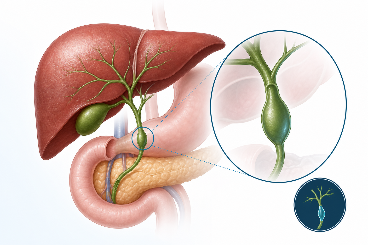
A choledochal cyst is an abnormal widening of part of the bile duct, the channel that carries bile from the liver toward the intestine. This change can affect bile flow and may lead to repeated inflammation, pain, or infection over time. Early specialist assessment helps define the extent of the cyst and supports timely treatment planning.
Causes and Risk Factors
Choledochal cyst is commonly related to congenital biliary tract development differences, meaning it may be present from birth even if symptoms appear later. Some patients are diagnosed in childhood, while others are identified in adolescence or adulthood during evaluation for abdominal symptoms or abnormal imaging findings. Prior biliary inflammation can further complicate presentation.
Symptoms
Symptoms can vary by age and cyst type. Patients may report upper abdominal pain, intermittent fever, nausea, jaundice (yellowing of eyes or skin), or episodes suggestive of cholangitis. Some individuals have mild or nonspecific symptoms, and the condition may be identified incidentally during ultrasound or other scans performed for a different concern.
Evaluation and Diagnosis
Evaluation usually starts with clinical history, physical examination, and baseline blood tests including liver function profile. Imaging plays a key role and may include abdominal ultrasonography and advanced cross-sectional biliary imaging to define duct anatomy and cyst involvement. A clear anatomical map guides surgical decision-making and perioperative planning.
Treatment Approach
Definitive management is typically individualized after imaging review and risk assessment. In many cases, treatment involves planned surgical management focused on removing diseased duct segments and restoring safe biliary drainage. The exact approach depends on cyst type, surrounding anatomy, current symptoms, and overall patient condition.
Recovery and Follow-up
Recovery planning includes pain control, nutrition progression, wound care guidance, and monitoring of liver-related parameters as advised by the treating team. Follow-up visits are important to review healing, discuss pathology where relevant, and confirm stable bile drainage on clinical and imaging review when needed. Patients should seek urgent care if fever, worsening pain, jaundice, or vomiting occurs.