
Hepatolithiasis
This page outlines symptoms, diagnosis, infection and obstruction concerns, treatment planning, and follow-up considerations to help patients and families understand when specialist consultation may be needed.
- Stone Disease
- Duct Imaging
- Surgical Planning
Condition Guide
Hepatolithiasis
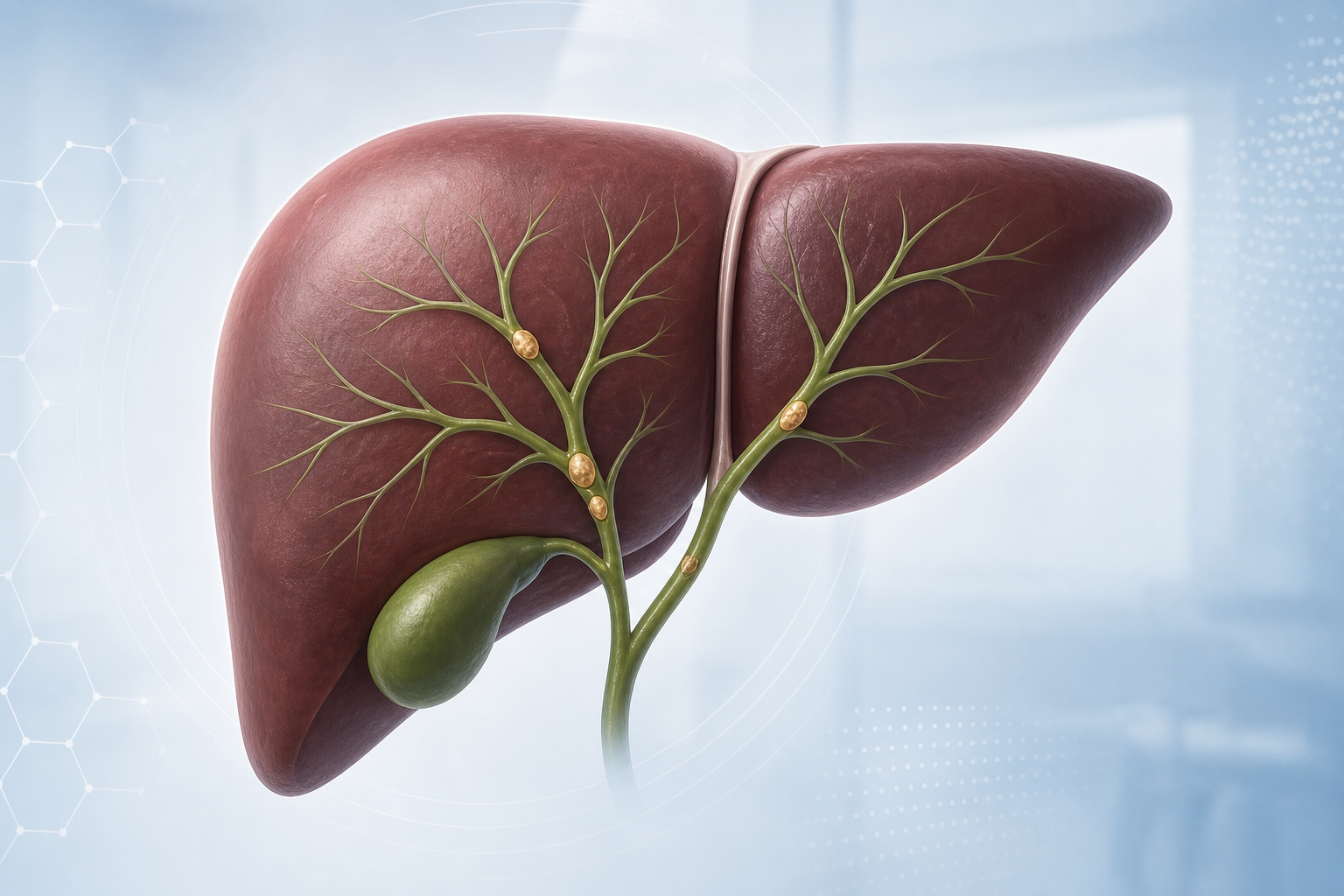
Hepatolithiasis refers to stones that form inside the bile ducts within the liver. This overview is designed to help patients and families understand the condition in simple terms, recognize when symptoms need attention, and review how diagnosis and treatment are planned.
Management is tailored to the pattern of stones, bile duct narrowing, and overall liver health. Early evaluation can reduce complications and support safer, more effective treatment decisions.
Overview
In hepatolithiasis, stones develop in the liver’s bile channels and can block normal bile flow. Over time, repeated blockage or infection may irritate bile ducts and surrounding liver tissue. The condition may remain quiet for periods, but flare-ups can occur and should be assessed promptly.
Causes and Risk Factors
Stone formation can be linked to bile stasis, recurrent biliary infection, bile duct narrowing, and changes in bile composition. Prior biliary surgery, anatomical variations, and delayed treatment of biliary disease may increase risk in some patients. Individual risk is best interpreted in clinical context.
Symptoms
Symptoms can include upper abdominal discomfort, fever, jaundice, nausea, reduced appetite, or repeated episodes of biliary infection. Some patients report intermittent symptoms that temporarily settle before returning. Persistent pain, fever, or yellowing of eyes/skin requires urgent medical review.
Evaluation and Diagnosis
Assessment usually begins with history, examination, and blood tests to evaluate liver function and inflammation. Imaging such as ultrasound, CT, or MRCP helps define stone location, duct dilation, and associated strictures. The final plan is based on imaging detail, symptom burden, and clinical stability.
Treatment Approach
Treatment may involve infection control, endoscopic or radiologic procedures to improve drainage, and selected surgical options when stones are extensive or recurrent. Care is typically individualized to clear obstruction, reduce recurrence risk, and preserve healthy liver tissue whenever possible.
Recovery and Follow-up
Recovery depends on the treatment pathway and baseline health. Follow-up generally focuses on symptom review, liver tests, and interval imaging when needed. Ongoing follow-up is important to detect recurrence early, support dietary and lifestyle guidance, and maintain long-term biliary health.
Need a personalized consultation for hepatolithiasis evaluation and treatment planning?
Book Appointment